Mouse TGFβ1 ELISA Kit
Size: 96T
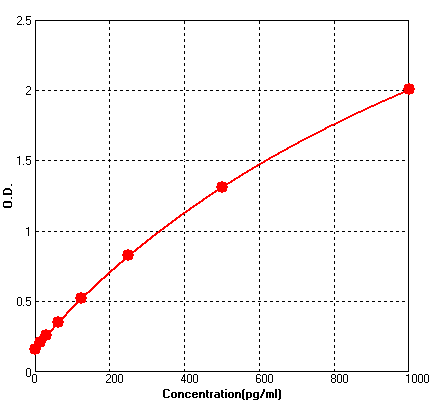
Range: 15.6 pg/ml - 1000 pg/ml
Sensitivity < 2 pg/ml
Application: For quantitative detection of TGFβ1 in mouse serum, body fluids, tissue lysates or cell culture supernatants.
--------------------------------------------------------------------------------------------------------------
Price: ---
Catalog No.: BEK1202
Size: 96T
Range: 15.6 pg/ml - 1000 pg/ml
Sensitivity < 2 pg/ml
Storage and Expiration: Store at 2-8℃ for 6 months, or at -20℃ for 12 months.
Application: For quantitative detection of TGFβ1 in mouse serum, plasma, body fluids, tissue lysates or cell culture supernatants.
Activating Reagent: TGFβ1 is mostly contained as inactive form in samples, please activate it with acid solution if want to analyze its active form.
Activating Reagent A: 1N HCI: add 8.33ml of 12N HCI into 91.67ml of H2O.
Activating Reagent B: 1.2N NaOH/0.5M HEPES: add 12ml of 10N NaOH and 11.9g HEPES into 75ml of H2O, add H2O to adjust volume to 100ml
Introduction
Transforming growth factor beta 1 or TGFβ1 is a polypeptide member of the transforming growth factor beta superfamily of cytokines. In humans, TGFβ1 is encoded by the TGFB1 gene. This gene contains 7 exons and very large introns, maps to 19q13.1-q13.3. TGFβ1 acts synergistically with TGFA in inducing transformation. It also acts as a negative autocrine growth factor. The TGFB1 is directly involved in the pathogenesis of bone marrow reticulin fibrosis in hairy cell leukemia. The expression of TGFB1 in the early stages of DMD may be critical in initiating muscle fibrosis, and suggested that antifibrosis treatment might slow progression of the disease, increasing the utility of gene therapy.
Principle of the Assay
This kit was based on sandwich enzyme-linked immune-sorbent assay technology. Anti- TGFβ1 polyclonal antibody was pre-coated onto 96-well plates. And the biotin conjugated anti- TGFβ1 polyclonal antibody was used as detection antibodies. The standards, test samples and biotin conjugated detection antibody were added to the wells subsequently, and wash with wash buffer. Avidin-Biotin-Peroxidase Complex was added and unbound conjugates were washed away with wash buffer. TMB substrates were used to visualize HRP enzymatic reaction. TMB was catalyzed by HRP to produce a blue color product that changed into yellow after adding acidic stop solution. The density of yellow is proportional to the TGFβ1 amount of sample captured in plate. Read the O.D. absorbance at 450nm in a microplate reader, and then the concentration of TGFβ1 can be calculated.
Kit components
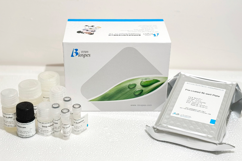
- One 96-well plate pre-coated with anti-mouse TGFβ1 antibody
- Lyophilized mouse TGFβ1 standards: 2 tubes (10 ng / tube)
- Sample / Standard diluent buffer: 30ml
- Biotin conjugated anti-mouse TGFβ1 antibody (Concentrated): 130μl. Dilution: 1:100
- Antibody diluent buffer: 12ml
- Avidin-Biotin-Peroxidase Complex (ABC) (Concentrated): 130μl. Dilution: 1:100
- ABC diluent buffer: 12ml
- TMB substrate: 10ml
- Stop solution: 10ml
- Wash buffer (25X): 30ml
- Activating Reagent A: 2 ml
- Activating Reagent B: 2 ml
Note: Reconstitute standards and test samples with Kit Component 3.
Material Required But Not Provided
- 37℃ incubator
- Microplate reader (wavelength: 450nm)
- Precise pipette and disposable pipette tips
- Automated plate washer
- ELISA shaker
- 1.5ml of Eppendorf tubes
- Plate cover
- Absorbent filter papers
- Plastic or glass container with volume of above 1L
