Mouse Renin-1 ELISA Kit
Size: 96T
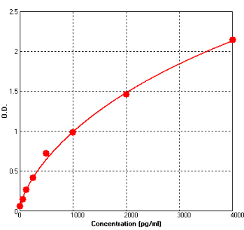
Range: 62.5 pg/ml-4000 pg/ml
Sensitivity < 10 pg/ml
Application: For quantitative detection of Renin-1 in mouse serum, plasma or cell culture supernates.
--------------------------------------------------------------------------------------------------------------
Price: $400.00
Catalog No.: BEK1252
Size: 96T
Range: 62.5 pg/ml-4000 pg/ml
Sensitivity < 10 pg/ml
Storage and Expiration: Store at 2-8℃ for 6 months, or at -20℃ for 12 months.
Application: For quantitative detection of Renin-1 in mouse serum, plasma or cell culture supernates.
Introduction
The secretion of renin from granules stored in renal juxtaglomerular cells plays a key role in blood pressure homeostasis. The synthesis and release of renin and the extent of granulation is regulated by several mechanisms including signaling from the macula densa, neuronal input, and blood pressure. Clark AF et al study indicate that expression of the Ren-1(d) gene is a prerequisite for the formation of storage granules, even though the related protein renin-2 is present in these mice, suggesting that renin-1(d) and renin-2 are secreted by distinct pathways in vivo. The synthesis of renin and other biologically active polypeptides in the granular convoluted tubule cells of the mouse submandibular gland (SMG) is regulated by androgen and thyroid hormones. The androgen and thyroid hormones influence levels of renin-1 in mouse SMG primarily by regulating the amount of renin-1 mRNA available for translation.
Principle of the Assay
This kit was based on sandwich enzyme-linked immune-sorbent assay technology. Anti-Renin-1 monoclonal antibody was pre-coated onto 96-well plates. And the biotin conjugated anti-Renin-1 polyclonal antibody was used as detection antibodies. The standards, test samples and biotin conjugated detection antibody were added to the wells subsequently, and wash with wash buffer. Avidin-Biotin-Peroxidase Complex was added and unbound conjugates were washed away with wash buffer. TMB substrates were used to visualize HRP enzymatic reaction. TMB was catalyzed by HRP to produce a blue color product that changed into yellow after adding acidic stop solution. The density of yellow is proportional to the Renin-1 amount of sample captured in plate. Read the O.D. absorbance at 450nm in a microplate reader, and then the concentration of Renin-1 can be calculated.
Kit components
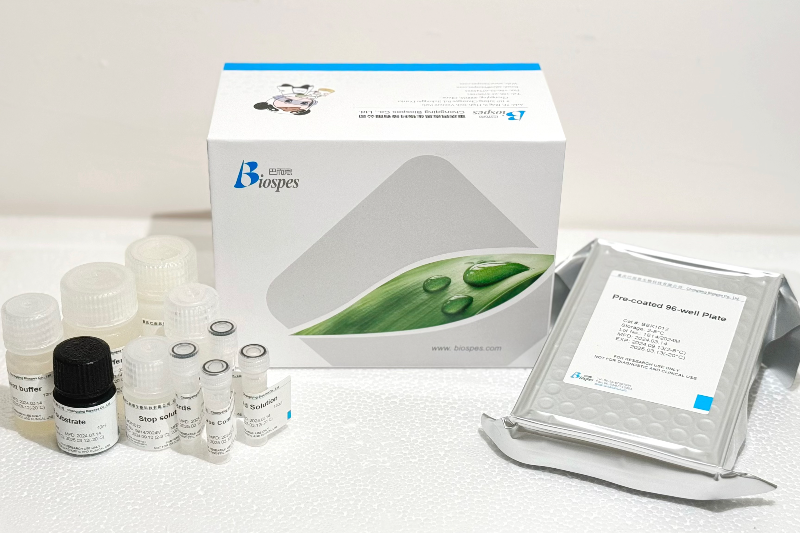
- One 96-well plate pre-coated with anti-mouse Renin-1 antibody
- Lyophilized Renin-1 standards: 2 tubes (10 ng / tube)
- Sample / Standard diluent buffer: 30 ml
- Biotin conjugated anti-mouse Renin-1 antibody (Concentrated): 130 μl. Dilution: 1:100
- Antibody diluent buffer: 12 ml
- Avidin-Biotin-Peroxidase Complex (ABC) (Concentrated): 130 μl. Dilution: 1:100
- ABC diluent buffer: 12 ml
- TMB substrate: 10 ml
- Stop solution: 10 ml
- Wash buffer (25X): 30 ml
Note: Reconstitute standards and test samples with Kit Component 3.
Material Required But Not Provided
- 37℃ incubator
- Microplate reader (wavelength: 450nm)
- Precise pipette and disposable pipette tips
- Automated plate washer
- ELISA shaker
- 1.5ml of Eppendorf tubes
- Plate cover
- Absorbent filter papers
- Plastic or glass container with volume of above 1L

