Human TNFα ELISA Kit
Size: 96T
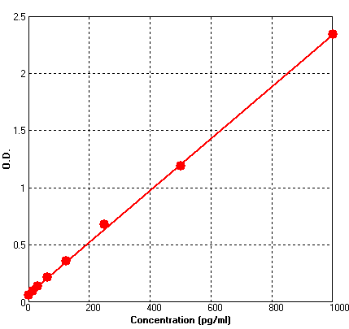
Range: 15.6 pg/ml-1000 pg/ml
(Body fluids, tissue lysates or cell culture supernates)
7.8 pg/ml-500 pg/ml (Human serum)
Sensitivity < 1 pg/ml
Application: For quantitative detection of TNFα in Human serum, body fluids, tissue lysates or cell culture supernatants.
--------------------------------------------------------------------------------------------------------------
Price: $320.00
Catalog No.: BEK1212
Size: 96T
Range: 15.6 pg/ml-1000 pg/ml (Body fluids, tissue lysates or cell culture supernates)
7.8 pg/ml-500 pg/ml (Human serum)
Sensitivity < 1 pg/ml
Storage and Expiration: Store at 2-8℃ for 6 months, or at -20℃ for 12 months.
Application: For quantitative detection of TNFα in Human serum, body fluids, tissue lysates or cell culture supernatants.
Introduction
Tumor necrosis factor, formerly known as tumor necrosis factor-alpha or TNF-α, is a cytokine involved in systemic inflammation and is a member of a group of cytokines that stimulate the acute phase reaction. It was originally identified in Human serum after injection with Mycobacterium bovis strain bacillus Calmette-Guerin (BCG) and endotoxin. TNF is primarily produced as a 212-amino acid-long type II transmembrane protein arranged in stable homotrimers. The human TNF gene maps to chromosome 6p21.3, spans about 3 kilobases and contains 4 exons. The primary role of TNF is in the regulation of immune cells. Dysregulation of TNF production has been implicated in a variety of human diseases, including Alzheimer's disease, cancer, major depression, and inflammatory bowel disease (IBD).
Principle of the Assay
This kit was based on sandwich enzyme-linked immune-sorbent assay technology. Anti- TNFα polyclonal antibody was pre-coated onto 96-well plates. And the biotin conjugated anti- TNFα polyclonal antibody was used as detection antibodies. The standards, test samples and biotin conjugated detection antibody were added to the wells subsequently, and wash with wash buffer. Avidin-Biotin-Peroxidase Complex was added and unbound conjugates were washed away with wash buffer. TMB substrates were used to visualize HRP enzymatic reaction. TMB was catalyzed by HRP to produce a blue color product that changed into yellow after adding acidic stop solution. The density of yellow is proportional to the TNFα amount of sample captured in plate. Read the O.D. absorbance at 450nm in a microplate reader, and then the concentration of TNFα can be calculated.
Kit components
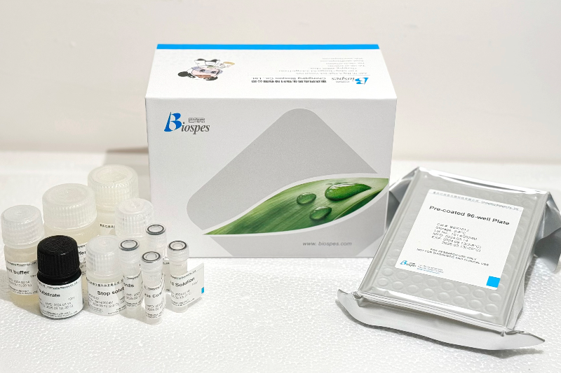
- One 96-well plate pre-coated with anti-Human TNFα antibody
- Lyophilized Human TNFα standards: 2 tubes (10 ng / tube)
- Sample / Standard diluent buffer: 30ml
- Biotin conjugated anti-Human TNFα antibody (Concentrated): 130μl. Dilution: 1:100
- Antibody diluent buffer: 12ml
- Avidin-Biotin-Peroxidase Complex (ABC) (Concentrated): 130μl. Dilution: 1:100
- ABC diluent buffer: 12ml
- TMB substrate: 10ml
- Stop solution: 10ml
- Wash buffer (25X): 30ml
Note: Reconstitute standards and test samples with Kit Component 3.
Material Required But Not Provided
- 37℃ incubator
- Microplate reader (wavelength: 450nm)
- Precise pipette and disposable pipette tips
- Automated plate washer
- ELISA shaker
- 1.5ml of Eppendorf tubes
- Plate cover
- Absorbent filter papers
- Plastic or glass container with volume of above 1L
